Скачать с ютуб Pathophysiology and Imaging in ARDS | Medvarsity в хорошем качестве
Pathophysiology and Imaging in ARDS | Medvarsity
2 года назад
Скачать бесплатно Pathophysiology and Imaging in ARDS | Medvarsity в качестве 4к (2к / 1080p)
У нас вы можете посмотреть бесплатно Pathophysiology and Imaging in ARDS | Medvarsity или скачать в максимальном доступном качестве, которое было загружено на ютуб. Для скачивания выберите вариант из формы ниже:
Загрузить музыку / рингтон Pathophysiology and Imaging in ARDS | Medvarsity в формате MP3:
Если кнопки скачивания не
загрузились
НАЖМИТЕ ЗДЕСЬ или обновите страницу
Если возникают проблемы со скачиванием, пожалуйста напишите в поддержку по адресу внизу
страницы.
Спасибо за использование сервиса savevideohd.ru
Pathophysiology and Imaging in ARDS | Medvarsity
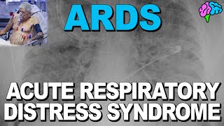
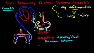
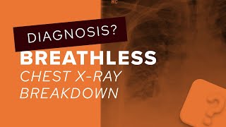
To know more or find more such videos please visit https://assimilate.in/ Acute Respiratory Distress Syndrome (ARDS) represents a stereotypical response to various etiologies. It progresses through several stages, beginning with alveolar-capillary damage, followed by a proliferative phase marked by improved lung function and healing, and finally a fibrotic phase signaling the end of the acute disease process. Focused changes on chest radiographs may be visible in patients with direct pulmonary insults. The initial radiograph in non-direct insults may be nonspecific or similar to congestive heart failure with mild effusions. Following that, interstitial pulmonary edema with diffuse infiltrates develops. As the disease progresses, the typical bilateral diffuse alveolar and reticular opacities appear. On a Computed Tomographic (CT) scan of the chest, the most prominent features of ARDS are diffuse consolidation with air bronchograms, bullae, pleural effusions, pneumomediastinum, and pneumothoraxes. Lung cysts of various sizes and numbers may appear later in the disease. A CT scan can detect ARDS complications as well as those related to catheter and tube placement, such as pneumothorax, pneumomediastinum, focal pneumonia, catheter malposition, and pulmonary infarction. Dr. Raymond Dominic Savio, discusses in detail about the pathophysiology and imaging in acute respiratory distress syndrome.
Comments