Скачать с ютуб Myoglobin and Hemoglobin (Compare and Contrast) в хорошем качестве
Myoglobin and Hemoglobin (Compare and Contrast)
10 лет назад
Скачать бесплатно Myoglobin and Hemoglobin (Compare and Contrast) в качестве 4к (2к / 1080p)
У нас вы можете посмотреть бесплатно Myoglobin and Hemoglobin (Compare and Contrast) или скачать в максимальном доступном качестве, которое было загружено на ютуб. Для скачивания выберите вариант из формы ниже:
Загрузить музыку / рингтон Myoglobin and Hemoglobin (Compare and Contrast) в формате MP3:
Если кнопки скачивания не
загрузились
НАЖМИТЕ ЗДЕСЬ или обновите страницу
Если возникают проблемы со скачиванием, пожалуйста напишите в поддержку по адресу внизу
страницы.
Спасибо за использование сервиса savevideohd.ru
Myoglobin and Hemoglobin (Compare and Contrast)
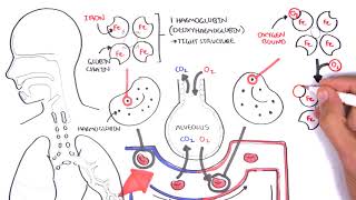
Moof's Medical Biochemistry Video Course: http://moof-university.thinkific.com/... NOTE: Correction: The histidine that anchors the heme group is the PROXIMAL* histidine, while the histidine that helps O2 bind is the DISTAL* histidine. In the video, I mistakenly switched that. My apologies. For Related Practice Problems with Worked Video Solutions on Protein Structure and Function, visit courses.moofuniversity.com. In this video, I discuss the structures, functions, and key characteristics of myoglobin and hemoglobin, as well as compare and contrast the two. Myoglobin (Mb) is a globular protein in muscle cells that functions in binding oxygen arriving to the muscles from the blood so as to deliver said oxygen to the muscle cells. Myoglobin has a single polypeptide chain with 8 alpha helices and no beta sheets. Since it has only one polypeptide chain, its highest level of protein structure is tertiary. It has one heme group (a prosthetic group) with an iron ion. It has two histidine residues in its interior, one of which is the proximal histidine, which anchors the heme group, while the other histidine residue, the distal histidine, helps myoglobin bind oxygen by reducing the binding affinity of carbon monoxide. Myoglobin’s oxygen binding curve is hyperbolic, characteristic of non-allosteric proteins. Hemoglobin (Hb) is a globular protein in red blood cells that functions in taking oxygen from the lungs to the tissues (including muscle tissue) via the bloodstream. Hemoglobin has quaternary structure, as it is made up of 4 polypeptide subunits, two alpha subunits, two beta subunits, each of which can bind one oxygen molecule (each subunit has a heme group). Hemoglobin displays cooperativity, a form of allosteric regulation; essentially, once one oxygen molecule is bound to one subunit of hemoglobin, the affinity for oxygen at the other subunits increases. It becomes easier for each successive oxygen molecule to bind hemoglobin. Hemoglobin’s oxygen binding curve is sigmoidal, characteristic of allosteric proteins. Between the two, Myoglobin has a higher affinity for oxygen than Hemoglobin. This can be see in their oxygen binding curves (shown in the video). This makes intuitive sense, though. If myoglobin functions by taking oxygen from hemoglobin in the blood to give to the muscle cells, then it MUST have a higher affinity for oxygen than hemoglobin. Otherwise, myoglobin wouldn’t be able to take the oxygen away from hemoglobin, and the muscle cells wouldn’t be able to be supplied with oxygen. NOTE: Correction: The histidine that anchors the heme group is the PROXIMAL* histidine, while the histidine that helps O2 bind is the DISTAL* histidine. In the video, I mistakenly switched that. My apologies. For a suggested viewing order of the videos, information on tutoring, personalized video solutions, and an opportunity to support Moof University financially, visit MoofUniversity.com, and follow Moof University on the different social media platforms. Don't forget to LIKE, COMMENT, and SUBSCRIBE: http://www.youtube.com/subscription_c... SUPPORT MOOF UNIVERSITY: http://www.moofuniversity.com/support... BUY A T-SHIRT https://shop.spreadshirt.com/moofuniv... INFORMATION ABOUT TUTORING AND PERSONALIZED VIDEO SOLUTIONS: http://www.moofuniversity.com/tutoring/ INSTAGRAM: / moofuniversity FACEBOOK: / 1554858934727545 TWITTER: / moofuniversity
Comments