Скачать с ютуб Clavicle Bone: Introduction, Anatomy, Function, Injuries and Treatment. в хорошем качестве
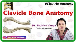
Clavicle Bone: Introduction, Anatomy, Function, Injuries and Treatment.
1 год назад
Скачать бесплатно Clavicle Bone: Introduction, Anatomy, Function, Injuries and Treatment. в качестве 4к (2к / 1080p)
У нас вы можете посмотреть бесплатно Clavicle Bone: Introduction, Anatomy, Function, Injuries and Treatment. или скачать в максимальном доступном качестве, которое было загружено на ютуб. Для скачивания выберите вариант из формы ниже:
Загрузить музыку / рингтон Clavicle Bone: Introduction, Anatomy, Function, Injuries and Treatment. в формате MP3:
Если кнопки скачивания не
загрузились
НАЖМИТЕ ЗДЕСЬ или обновите страницу
Если возникают проблемы со скачиванием, пожалуйста напишите в поддержку по адресу внизу
страницы.
Спасибо за использование сервиса savevideohd.ru
Clavicle Bone: Introduction, Anatomy, Function, Injuries and Treatment.
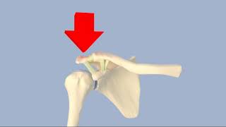
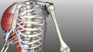
. Chapters 0:00 Introduction 0:42 Anatomy 2:03 Functions of Clavicle Bone 2:34 Injuries from Clavicle Bone 3:27 Treatment for Clavicle Bone The clavicle, or collarbone, is a slender, S-shaped long bone approximately 6 inches (15 cm) long[1] that serves as a strut between the shoulder blade and the sternum (breastbone). There are two clavicles, one on the left and one on the right. The clavicle is the only long bone in the body that lies horizontally. Together with the shoulder blade, it makes up the shoulder girdle. It is a touchable bone, and in people who have less fat in this region, the location of the bone is clearly visible, as it creates a bulge in the skin. It receives its name from the Latin clavicula ("little key"), because the bone rotates along its axis like a key when the shoulder is abducted. The clavicle is the most commonly fractured bone. It can easily be fractured by impacts to the shoulder from the force of falling on outstretched arms or by a direct hit.[2]The collarbone is a thin doubly curved long bone that connects the arm to the trunk of the body. Located directly above the first rib, it acts as a strut to keep the scapula in place so that the arm can hang freely. At its rounded medial end (sternal end), it articulates with the manubrium of the sternum (breastbone) at the sternoclavicular joint. At its flattened lateral end (acromial end), it articulates with the acromion, a process of the scapula (shoulder blade), at the acromioclavicular joint. Clavicula inf.jpg Clavicula sup.jpg Right clavicle—from below, and from above Gray200.png Gray201.png Left clavicle—from above, and from below The rounded medial region (sternal region) of the shaft has a long curve laterally and anteriorly along two-thirds of the entire shaft. The flattened lateral region (acromial region) of the shaft has an even larger posterior curve to articulate with the acromion of the scapula. The medial region is the longest clavicular region as it takes up two-thirds of the entire shaft. The lateral region is both the widest clavicular region and thinnest clavicular region. The lateral end has a rough inferior surface that bears a ridge, the trapezoid line, and a slight rounded projection, the conoid tubercle (above the coracoid process). These surface features are attachment sites for muscles and ligaments of the shoulder. It can be divided into three parts: medial end, lateral end, and shaft. Medial end The medial end is also known as the sternal end. It is quadrangular and articulates with the clavicular notch of the manubrium of the sternum to form the sternoclavicular joint. The articular surface extends to the inferior aspect for articulation with the first costal cartilage. Lateral end The lateral end is also known as the acromial end. It is flat from above downward. It bears a facet that articulates with the shoulder to form the acromioclavicular joint. The area surrounding the joint gives an attachment to the joint capsule. The anterior border is concave forward and the posterior border is convex backward. Shaft The shaft is divided into two main regions, the medial region, and the lateral region. The medial region is also known as the sternal region, it is the longest clavicular region as it takes up two-thirds of the entire shaft. The lateral region is also known as the acromial region, it is both the widest clavicular region and thinnest clavicular region. 3D model of the clavicle Lateral region of the shaft The lateral region of the shaft has two borders and two surfaces. the anterior border is concave forward and gives origin to the deltoid muscle. the posterior border is convex and gives attachment to the trapezius muscle. the inferior surface has a ridge called the trapezoid line and a tubercle; the conoid tubercle for attachment with the trapezoid and the conoid ligament, part of the coracoclavicular ligament that serves to connect the collarbone with the coracoid process of the scapula. Development The collarbone is the first bone to begin the process of ossification (laying down of minerals onto a preformed matrix) during development of the embryo, during the fifth and sixth weeks of gestation. However, it is one of the last bones to finish ossification at about 21–25 years of age. Its lateral end is formed by intramembranous ossification while medially it is formed by endochondral ossification. It consists of a mass of cancellous bone surrounded by a compact bone shell. The cancellous bone forms via two ossification centres, one medial and one lateral, which fuse later on. The compact forms as the layer of fascia covering the bone stimulate the ossification of adjacent tissue. The resulting compact bone is known as a periosteal collar.
Comments