Скачать с ютуб Transcatheter Aortic Valve Implantation (TAVI) в хорошем качестве
Transcatheter Aortic Valve Implantation (TAVI)
11 месяцев назад
Скачать бесплатно Transcatheter Aortic Valve Implantation (TAVI) в качестве 4к (2к / 1080p)
У нас вы можете посмотреть бесплатно Transcatheter Aortic Valve Implantation (TAVI) или скачать в максимальном доступном качестве, которое было загружено на ютуб. Для скачивания выберите вариант из формы ниже:
Загрузить музыку / рингтон Transcatheter Aortic Valve Implantation (TAVI) в формате MP3:
Если кнопки скачивания не
загрузились
НАЖМИТЕ ЗДЕСЬ или обновите страницу
Если возникают проблемы со скачиванием, пожалуйста напишите в поддержку по адресу внизу
страницы.
Спасибо за использование сервиса savevideohd.ru
Transcatheter Aortic Valve Implantation (TAVI)
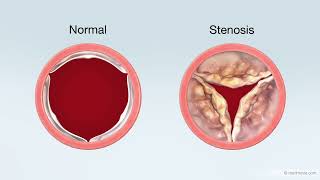
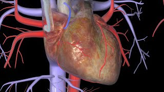
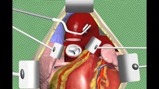
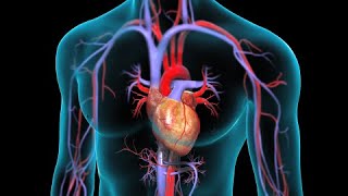
MEDICAL ANIMATION TRANSCRIPT: If you have a severe problem with the aortic valve in your heart, your doctor may recommend a transcatheter aortic valve implantation, or TAVI. It's also known as transcatheter aortic valve replacement, or TAVR. Your aortic valve is one of the four valves in your heart. It lies between your left ventricle, or lower left chamber, and the large blood vessel supplying oxygen rich blood to your body, called the aorta. When your left ventricle contracts during each heartbeat, it pushes blood through three flexible cup-like leaflets that make up your aortic valve. When your left ventricle relaxes, your aortic valve closes to prevent blood from flowing back into your left ventricle. As you get older, your aortic valve may develop deposits of calcium that cause your leaflets to get thicker and less flexible. In this condition, your leaflets may not open fully. This condition, called calcific aortic valve stenosis, creates a narrowed opening that reduces the amount of blood flowing into your aorta and out to your body. As a result, the wall of your left ventricle thickens because it has to work harder to pump blood through the narrowed valve opening. At the beginning of your procedure, your doctor will make a small incision in your groin. After creating an opening in a blood vessel called the femoral artery your doctor will insert a flexible tube, called an introducer sheath. Through the sheath, your doctor will insert a flexible guidewire into your femoral artery. The guidewire will be passed through your femoral artery all the way up to your aorta. Then, the guidewire will be pushed through the opening in your aortic valve and into your left ventricle. Your doctor will use the guidewire to pass a flexible tube called a catheter through your aortic valve. A balloon on the tip of the catheter will be inflated to widen the opening in your aortic valve, to push the valve leaflets to the sides. After deflating the balloon, your doctor will remove the catheter. The replacement valve consists of a tissue valve surrounded by a wire mesh called a stent. The replacement valve will be compressed and placed on the tip of another catheter. This catheter will be inserted through your aortic valve. When the catheter reaches the opening in your aortic valve, your surgeon will inflate a balloon underneath the replacement valve to expand it and the stent. After placement of the new aortic valve, your doctor will deflate the balloon. The stent will support and secure the replacement valve in place. The catheter and guidewire will be removed. At the end of the procedure, the introducer sheath in your groin will be removed and your skin incision will be closed. You will have another incision on the other side of your groin through which instruments were inserted to monitor your heart during the procedure. This skin incision will also be closed. To find out more about transcatheter aortic valve implantation, talk to your healthcare practitioner. #TAVI #TAVR #AorticValveReplacement ANH23268
Comments